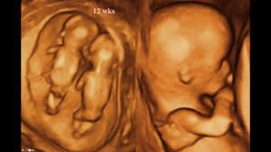
A fetal anomaly scan (or anatomy scan) is a detailed, second-trimester ultrasound that examines your baby’s organs, skeleton, brain, and heart. Typically performed between 18 and 22 weeks of pregnancy, it ensures the fetus is growing normally and screens for structural birth defects.
Why Is It Performed?
The primary goal is to provide a comprehensive "head-to-toe" check of your baby's development. Specifically, the scan evaluates: Internal Organs: Checks the brain, heart, stomach, kidneys, and bowel for structural abnormalities. Physical Structure: Examines the face (including lips for clefts), spine, limbs, and fingers/toes.
Growth Metrics:
Takes precise measurements of the head, abdomen, and thigh bones to ensure they match gestational age. Pregnancy Environment: Assesses the amniotic fluid volume and the position of the placenta. What to Expect During the Procedure The Process: Similar to standard ultrasounds, you will lie on an exam table while a technician applies a warm gel to your abdomen and uses a handheld transducer to send sound waves into the womb. Duration: It is highly detailed and typically takes 30 to 45 minutes.
Knowing the Sex:
In most cases, if you wish to know the sex of the baby, the sonographer can check this for you during the scan. Benefits and Limitations While this scan is a vital tool for prenatal care, it is important to understand its capabilities.
Preparation:
Detecting a condition early allows parents and medical teams to plan for specialized delivery or medical interventions immediately after birth.
Limitations:
Not all congenital conditions can be detected via ultrasound. Some structural defects may be too minor to visualize, develop later in pregnancy, or be obscured by the baby's position.
For more localized or specific guidelines, you can review the NHS 20-week Screening Scan overview or consult your maternal-fetal medicine specialist for regional testing protocols.
About the Doctor

Dr. Naren Satya
Consultant Radiologist
Naren Ultrasound and Fetal Medicine Center, Visakhapatnam
Related Topics